AI Projects
WCRAI - The Wilhelm Conrad Röntgen Artificial Intelligence
Introduction
Pneumonia is a lung condition that presents significant challenges in its diagnosis and treatment, being a major cause of morbidity and mortality worldwide. The interpretation of radiographic images plays a crucial role in the detection of this disease, but the subtle and variable nature of the radiographic manifestations of pneumonia can lead to uncertain diagnoses and delays in appropriate treatment. In this context, artificial intelligence (AI) emerges as a revolutionary tool that promises a significant improvement in the accuracy and efficiency of radiological diagnosis.The WCRAI project (Wilhelm Conrad Röntgen Artificial Intelligence), named in honor of the radiography pioneer, seeks to address these challenges through the implementation of advanced machine learning and computer vision techniques. The goal is to develop an AI algorithm capable of analyzing X-ray images to not only detect the presence of pneumonia but also differentiate between its various types, such as viral and bacterial pneumonia, with a high degree of accuracy and reliability.
This project not only aspires to improve patient outcomes through faster and more accurate diagnoses but also aims to reduce the burden on radiologists, allowing them to focus on more complex cases and critical clinical decision-making. With the implementation of WCRAI, the door is opened to a future where technology and medicine merge to offer higher quality healthcare, accessible to a broader population, and with the possibility of applying the same technology to other pathologies and aspects of radiodiagnosis.
The commitment to innovation and excellence in healthcare is the driving force of this project, fueled by the conviction that AI technology has the potential not only to transform radiology but to significantly improve the lives of patients around the world. With a vision directed towards the future and a focus on sustainable and scalable solutions, WCRAI positions itself at the forefront of the intersection between technology and medicine.
Project goals
The WCRAI project focuses on the development and refinement of a state-of-the-art artificial intelligence system specifically designed for the accurate detection and classification of pneumonia from X-ray images. With the goal of significantly improving the quality and speed of radiological diagnosis, the project establishes the following specific objectives:1. Improvement of diagnostic accuracy: Refine the algorithm's ability to identify and differentiate between viral and bacterial pneumonia, minimizing false positives and negatives. This will allow more specific and timely treatment for patients, which is crucial in the efficient management of the disease.
2. Reduction of diagnostic time: Speed up the diagnostic process through the automatic and real-time interpretation of X-ray images, facilitating a quick response that is essential in cases of high urgency.
3. Support in clinical decision-making: Provide healthcare professionals with an additional tool for the evaluation of complex cases, contributing to evidence-based decision-making and reinforcing confidence in radiological diagnoses.
4. Training and continuous learning of the algorithm: Implement deep learning methods that allow the system to continuously improve its performance through the analysis of new data, ensuring constant evolution in its accuracy and reliability.
5. Optimization of radiological workflow: Integrate the AI system within the existing radiological workflow to improve operational efficiency, reduce the workload of radiologists, and allow them to focus on cases that require their specialized expertise.
6. Democratization of access to quality diagnosis: Facilitate access to high-quality diagnoses in regions with a shortage of specialists, contributing to equity in healthcare and supporting overburdened health systems.
7. Extension and scalability: Develop a system that can be easily adapted for the diagnosis of other medical conditions, demonstrating the flexibility and scalability of the AI algorithm in various clinical applications.
Each of these objectives contributes to the main mission of the WCRAI project: to revolutionize the field of radiology and improve patient care through artificial intelligence. With a firm commitment to responsible and ethical innovation, the project seeks to establish a new standard in the accuracy and efficiency of medical diagnosis.
Methodology
The WCRAI project relies on a robust and systematic methodology to develop an artificial intelligence algorithm capable of diagnosing and differentiating types of pneumonia through chest radiographic images. The methodological steps are as follows:1. Selection and preparation of data:
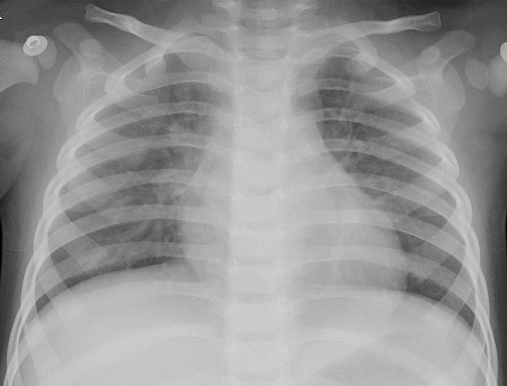
Radiographic images were obtained from Paul Mooney's "Chest X-Ray Images (Pneumonia)" database on Kaggle, which includes 5.388 chest X-rays previously labeled as "no pneumonia", "pneumonia", "viral pneumonia", and "bacterial pneumonia". All images were meticulously reviewed and validated to ensure the quality and accuracy of the labels.
2. Image processing:
To optimize the model training process, the images were resized to a uniform size, allowing more efficient analysis and reducing the variability that could introduce noise into the learning model.
3. Model training:
The algorithm training was divided into two essential phases:
- Phase 1: A model was trained to distinguish between "no pneumonia" and "pneumonia" images.
- Phase 2: Subsequently, a second model was trained to differentiate between "bacterial pneumonia" and "viral pneumonia".
4. User interface development:
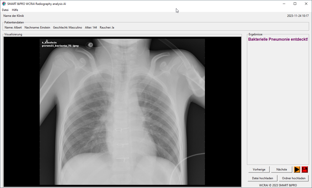
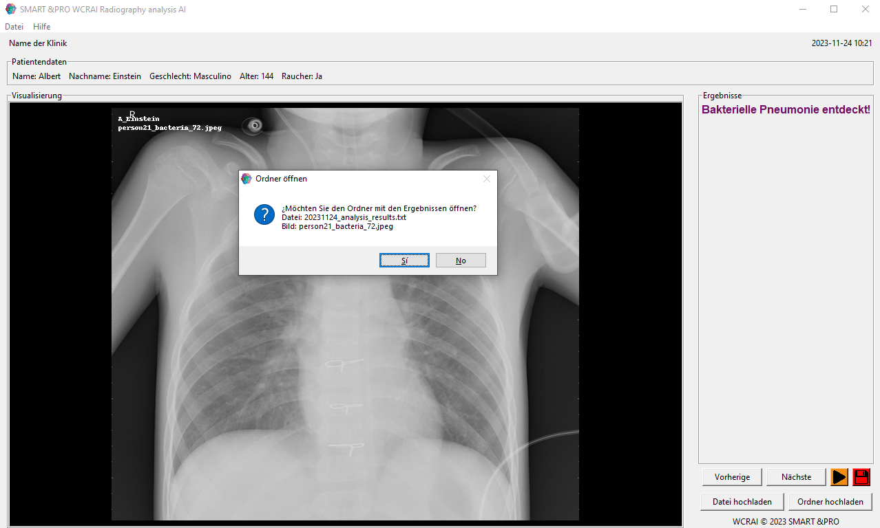
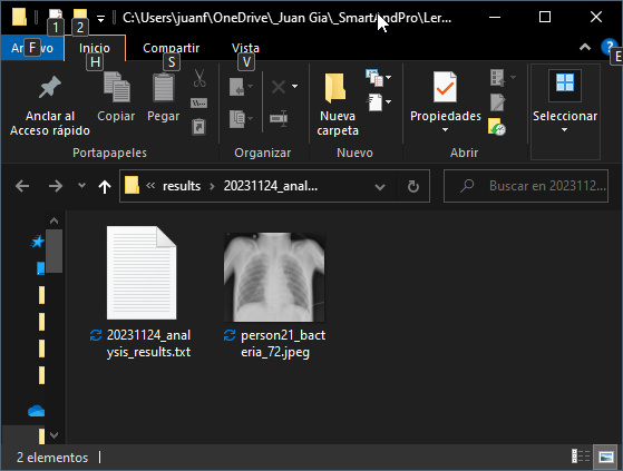
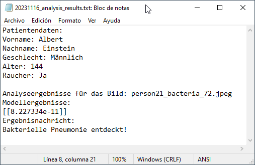
An intuitive user interface was programmed using Tkinter. This interface first loads an image or a folder of images and patient data. The interface allows viewing the image or images one by one, showing both the name of the file being viewed and the name of the folder in which it is located. The user decides when to run the algorithm. This proceeds with the execution of Phase 1 to determine the presence of pneumonia. If the result is positive, then the Phase 2 algorithm is automatically activated, providing the user with a more detailed diagnosis between "bacterial pneumonia" or "viral pneumonia". Finally, the user can automatically save the analysis information and later view the folder and its contained files. All the analysis information is recorded in a plain text file which provides the user with the following information: Patient data, analysis date, image name, name of the containing folder, algorithm accuracy result, and the final message shown to the user as a result of the analysis.
5. Implementation and technical evaluation:
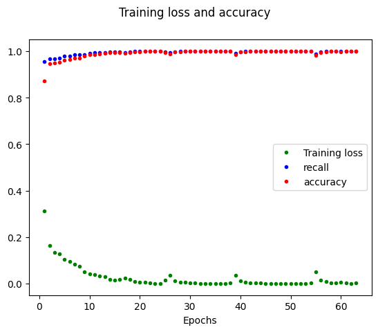
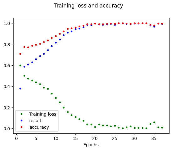
A set of deep learning tools, including TensorFlow and Keras, were used for the construction and training of the neural convolution models. Techniques such as data splitting into training and test sets, cross-validation, and callbacks to avoid overfitting were employed. In addition, the model's performance was monitored using metrics such as training loss, accuracy, and recall, and its performance was visualized through evolution graphs and confusion matrices.
6. Model validation:
The model's performance was evaluated by comparing the model's predictions with the true labels of the test data. The true positive rate was calculated and the model was adjusted to maximize this metric, which is especially critically important in the medical context where false negatives can have serious consequences.
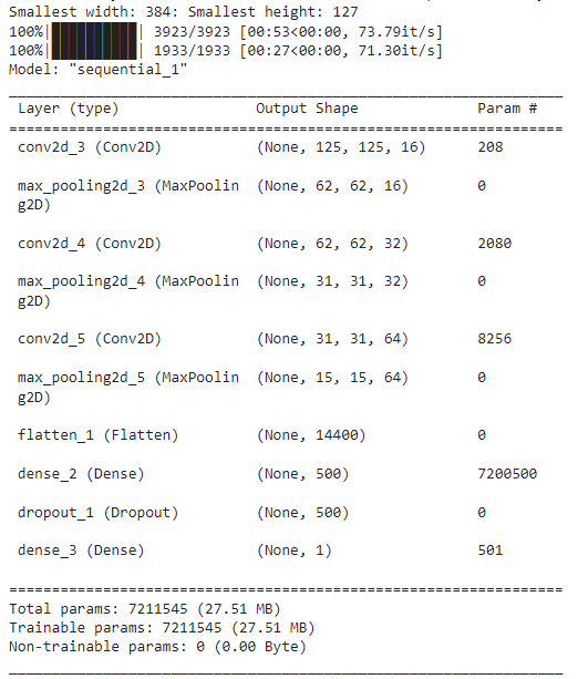
Arquitectura del Modelo de Red Neuronal Convolucional:
The AI model developed in the WCRAI project is composed of a sequence of convolution and max-pooling layers, followed by dense layers and a dropout layer to prevent overfitting. Specifically, the network begins with a convolutional layer (Conv2D) with a 2x2 kernel size and a ReLU activation function, designed to extract low-level features from the input images. This layer is followed by a max-pooling layer that reduces data dimensionality to improve computational efficiency and prevent overfitting.The network continues to deepen with additional convolutional and max-pooling layers, increasing in the number of filters, allowing the model to learn more complex and abstract patterns as it progresses through the architecture. After the convolutional and pooling layers, the network flattens the multidimensional data to process it through dense layers, culminating in an output layer with a single unit and a sigmoid activation function for binary classification.
The inclusion of a dropout layer before the output layer acts as a regularization technique, randomly discarding a fraction of the connections during training to make the network more robust to variations in the input data.
The model is characterized by a significant number of trainable parameters, indicating its capacity to learn a wide variety of features from chest radiographic images. However, the size and complexity of the model also underline the need for a substantial and diverse training dataset to effectively train it.
Development of the WCRAI algorithm
The development of the WCRAI algorithm constitutes the technical core of the project and is a testament to the practical application of artificial intelligence in the field of radiology. The process of creating this advanced system is divided into clearly defined stages, each with its specific focus on improving the accuracy and usability of pneumonia diagnosis.Technology selection and architecture design:
The first stage of development involved selecting an appropriate technological framework. TensorFlow was chosen for its flexibility and robustness, as well as Keras for its high-level interface and ease of use. The architecture of the model was designed as a convolutional neural network (CNN), recognized for its effectiveness in computer vision and deep learning tasks.
Preprocessing and data augmentation:
A critical step in development was the preprocessing of X-ray images. The images were normalized and resized to ensure uniformity in the model's input. In addition, data augmentation techniques were implemented to artificially expand the training set, thus improving the model’s generalization.
Model programming and training:
The model's programming was carried out using Python, a high-level programming language with a wide range of support libraries for machine learning. The model training was conducted in two phases. In the first, images of 'non-pneumonia' and 'pneumonia' were differentiated; and in the second, 'pneumonia' images were classified into 'viral' and 'bacterial'. This stepped approach optimized the model's accuracy and efficiency.
User interface and integration testing:
Subsequently, a user interface (UI) was developed using Tkinter, a Python library for creating graphical interfaces. The UI allows intuitive interaction with the algorithm and facilitates the interpretation of its results. Extensive testing was conducted to ensure a smooth integration of the model with the UI, prioritizing the end-user experience.
Validation and Tine-Tuning:
Once the model was trained, its performance was validated using an independent test dataset. Performance metrics such as accuracy, sensitivity, and specificity were analyzed, and the model was fine-tuned based on these results.
Documentation and knowledge transfer:
The knowledge and experiences gained during the development of the algorithm were meticulously documented. This documentation serves as a valuable resource for a deep understanding of the system and facilitates future improvements and maintenance.
Results
The results phase of the WCRAI project focused on the comprehensive evaluation of the artificial intelligence model developed for the detection and classification of pneumonia. The results obtained are critically important, as they provide tangible validation of the algorithm's effectiveness and its applicability in real clinical settings.Model accuracy evaluation:
The model demonstrated high accuracy in identifying pneumonia in chest radiographic images. Specific metrics, including accuracy, sensitivity (recall), and the F-value, indicated that the algorithm has an excellent ability to distinguish between normal images and those indicative of pneumonia, as well as between viral and bacterial pneumonia.
Analysis of the confusion matrix:
The confusion matrices for each phase of the model revealed that true positives and true negatives constituted the majority of predictions, with a relatively low number of false positives and false negatives. This suggests that the model is highly reliable and minimizes the risks associated with misdiagnoses.
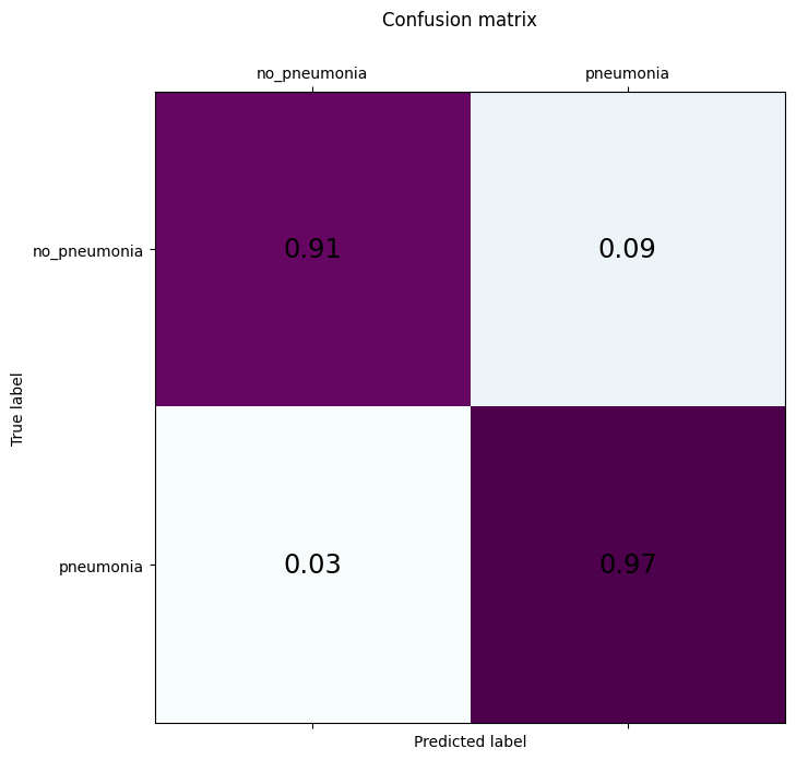
Accuracy Phase 1: - “no neumonia” = 0.91 % - “neumonia” = 0.97 % |
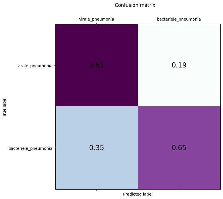
Accuracy Phase 2: - “virale neumonia” = 0.81 % - “bacteriale neumonia” = 0.65 % |
Comparison with established benchmarks:
When comparing the performance of the WCRAI model with current standards and other relevant studies, it was found that the system provided comparable or superior results, suggesting that it has the potential to significantly improve diagnostic accuracy in medical practice.
Results of the Fine-Tuning process:
Fine-tuning the model through the analysis of ROC and AUC curves resulted in an even more robust system, with an excellent balance between the rate of detection of true positive cases and the minimization of false alarms.
User feedback:
Feedback from other professionals who interacted with the model and user interface was largely very positive, highlighting the system's speed and ease of use, as well as the clarity of the presented results.
Clinical impact:
Analysis of the results indicated that the WCRAI model has the potential to play a significant role in improving diagnostic workflows for pneumonia, offering valuable decision support for physicians and ultimately contributing to more effective patient management.
Clinical implementation
The clinical implementation of the WCRAI algorithm represents a step forward in integrating artificial intelligence into everyday medical practice. This process encompasses not only the technical installation of the software but also a series of critical stages to ensure the successful adoption and operational effectiveness of the system.System integration:
The WCRAI algorithm was designed to be compatible with existing hospital information systems, facilitating a seamless integration. Special attention is paid to interoperability with PACS (Picture Archiving and Communication Systems) and EMR (Electronic Medical Records), allowing the AI system to become a synergistic part of the radiological workflow.
Staff training:
A comprehensive training program for radiologists and technicians was developed, ensuring that staff is fully informed on how to use the system. This includes handling the user interface, interpreting the results of the algorithm, and knowledge of response protocols in case of pneumonia detection.
Continuous support and maintenance:
A continuous technical support plan is established to resolve any technical issues or queries that may arise, thus ensuring maximum system availability and performance. This includes regular software maintenance and algorithm updates based on the latest advancements and research.
Impact assessment and continuous improvement:
Following implementation, a constant evaluation of the clinical impact of the WCRAI algorithm will be conducted. This evaluation measures not only diagnostic accuracy but also workflow efficiency and patient satisfaction. The results of these evaluations will inform the system's continuous improvements.
Clinical studies and publications:
To validate the clinical efficacy of the algorithm, rigorous clinical studies will be conducted, documented, and published. This not only serves to establish the system's validity but also to increase confidence in AI within the medical community.
Adherence to standards and regulations:
The entire implementation is carried out in strict adherence to medical standards and data privacy regulations, such as HIPAA in the United States, ensuring that patient safety and data confidentiality are paramount.
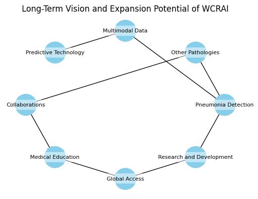
Future vision and expansion potential
The future vision of the WCRAI project extends beyond its current application in pneumonia diagnosis, projecting towards a wide range of possibilities and applications in the field of diagnostic medicine. Scalability and adaptability are inherent features of the system's design, allowing for continuous evolution and expansion.Expansion to other pathologies:
The WCRAI algorithm was built with a modular architecture that allows for the incorporation of new diagnostic modules. In the future, the algorithm is expected to expand its capability to include the detection and classification of other thoracic and pulmonary diseases, such as tuberculosis, lung cancer, and interstitial lung diseases.
Integration of multimodal data:
With a view towards a more holistic approach to diagnosis, the project envisions integrating data from multiple imaging modalities, such as computed tomography (CT) and magnetic resonance imaging (MRI), as well as clinical and laboratory data, to provide a more comprehensive and accurate analysis.
Development of predictive technology:
Long-term goals include advancing towards a system that not only diagnoses but also predicts disease progression and responds to treatments, using predictive analysis and deep learning techniques. This could have significant implications for treatment planning and disease monitoring.
Interdisciplinary collaborations:
Efforts are being made to foster collaborations with experts in other disciplines, such as epidemiology and genomics, to explore how patterns in radiological data can correlate with epidemiological trends or genetic markers, thus expanding the scope of WCRAI into research and public health.
Involvement in medical education:
The WCRAI system also has the potential to be an educational tool, helping to train future radiologists and technicians in image interpretation and the integration of AI into clinical practice.
Global adoption and equitable access:
A critically important vision is the democratization of access to advanced diagnostic technology. WCRAI strives to make diagnostic AI accessible worldwide, even in resource-limited regions, thus contributing to the reduction of healthcare disparities.
Continuous research and development:
The WCRAI project is committed to ongoing research and development, keeping the system at the forefront of technology and ensuring that it adapts and evolves with the latest advancements in AI and medicine.
Looking towards the future, WCRAI not only positions itself as a leading system in pneumonia diagnosis through AI but also as a pioneer in the field of computer-assisted diagnostic medicine, with the potential to profoundly transform the way healthcare professionals interact with technology and manage patient care.
Challenges and solutions
In the development and implementation of Artificial Intelligence in medical diagnosis, the WCRAI project has faced several significant challenges. Identifying and overcoming these obstacles has been crucial to ensuring the effectiveness and acceptance of the system.Image quality variability:
Variability in the quality and format of X-ray images can affect algorithm performance. To address this, a rigorous preprocessing process has been implemented to normalize and standardize images before introducing them into the system, ensuring that the model receives consistent data for analysis.
Data integration:
Integrating data from multiple sources and formats presents a technical challenge. Collaboration with EMR and PACS providers is sought to develop Application Programming Interfaces (APIs) that facilitate seamless data integration.
Overfitting:
Overfitting is a common risk in machine learning, where a model can memorize training data, losing the ability to generalize to new data. To combat this, techniques such as cross-validation and dropout have been used, and data augmentation is sought to expand and diversify the training dataset.
Adoption by medical professionals:
Resistance to adopting AI in clinical practice is another challenge. To overcome this barrier, seminars and educational workshops are being organized to demonstrate the functionality and benefits of the WCRAI system, and active feedback from professionals is sought to improve the interface and user experience.
Ethical and privacy standards:
Compliance with ethical standards and privacy regulations is paramount. Ensuring that the system complies with data privacy regulations such as HIPAA has been a priority, and transparency has been incorporated into the algorithm's decision-making process.
Scalability and maintenance:
Ensuring that the system can scale and be maintained over time is essential. The modular design of the WCRAI system and the focus on developing standardized APIs enable easy system updates and expansion.
International interoperability:
The diversity of international medical standards and practices can hinder global implementation. To address this, efforts are being made toward an adaptable system that can be configured according to local needs and regulations.
Ongoing technological advancement:
The rapid evolution of AI technology means that the system must be continuously updated. A commitment to ongoing research and development ensures that the WCRAI system remains at the forefront of technology.
Challenges and Solutions in the WCRAI Project Implementation
| Challenges | Solutions | |
|---|---|---|
| Variability in image quality | ➔ | Normalization and standardization |
| Data integration | ➔ | API development with EMR/PACS |
| Overfitting | ➔ | Cross-Validation and dropout |
| Adoption by medical professionals | ➔ | Educational seminars and workshops |
| Ethical and privacy standards | ➔ | Compliance with HIPAA |
| Scalability and maintenance | ➔ | Modular design and standardized APIs |
| International interoperability | ➔ | Configuration according to local norms |
| Continuous technological advancement | ➔ | Ongoing research and development |
Facing these challenges and finding appropriate solutions has been and continues to be an integral part of the WCRAI project process, and it remains essential to build a robust and reliable system that can withstand the rigorous demands of the medical environment
Future steps
The future steps for the WCRAI project are designed to ensure that the system not only remains relevant and effective in the face of technological advancements but also continues to meet the changing needs of healthcare professionals and patients. These steps focus on expansion, feedback integration, and continuous improvement.Expansion of the dataset:
The first step involves the ongoing collection and analysis of additional radiographic images with the aim of expanding and diversifying the dataset. This will enhance the model's ability to generalize and operate effectively in a broader and more diverse patient population.
Algorithm update and evolution:
In parallel, there are plans to periodically update the algorithm to incorporate the latest advances in machine learning and computer vision techniques. This will include experimenting with new models and neural network architectures to improve diagnostic accuracy and speed.
Integration of clinical feedback:
Efforts will be made to establish a mechanism for integrating feedback from end users into the system. The input of radiologists, medical imaging technicians, and other healthcare professionals using WCRAI will be crucial in guiding the development of additional features and user interface improvements.
Development of explanatory AI features:
To increase confidence in the clinical use of WCRAI, work will be done to develop features that provide clear and understandable explanations of the algorithm's decisions and results, making the AI more transparent and less like a "black box."
Promotion of inter-institutional collaborations:
Efforts will be made to establish partnerships with hospitals, universities, and research centers to promote clinical studies that can further validate the effectiveness of WCRAI and explore new clinical applications for the technology.
Promotion of awareness and education:
Efforts will be made to educate the medical community and the general public about the benefits of AI in medical diagnosis. This will include participation in conferences, publication of articles, and the creation of educational materials.
Regulatory assessments and compliance:
A critical step will be ongoing navigation of the regulatory landscape to ensure that WCRAI complies with all medical device regulations in the markets where it operates, including obtaining regulatory approvals where necessary.
Preparation for large-scale implementation:
Finally, strategies will be prepared for large-scale implementation of the system, including the development of infrastructure and technical support plans to facilitate widespread adoption of WCRAI in a variety of clinical settings.
Contact and project WCRAI references
Project main contact:
Juan García
CEO, AI Expert & AI Manager
SMART &PRO AI Services
[email protected]
Tel.: (+49) 162 5371628
Stühlingen (Germany)
Bibliographic references:
Neural architecture search for pneumonia diagnosis from chest X-rays,
Abhibha Gupta, Parth Sheth & Pengtao Xie, 2022, 10.1038/s41598-022-15341-0,
https://www.nature.com/articles/s41598-022-15341-0
Online resources:
- "Chest X-Ray images (Pneumonia)" por Paul Mooney en Kaggle: https://www.kaggle.com/datasets/paultimothymooney/chest-xray-pneumonia
- WCRAI_Präsentation.pdf
-
Paper publication date:
November 17, 2023
-
Code Website:
Github
* NOTICE: The code is protected by copyright. Its use is only allowed for educational purposes with the written permission of the owner. -
Paper download:
 Español
 English
 Deutsch
-
Logos:
Social Media:
AI Projects